
Evaluation of Si-Trap™ for Integrated Proteomic, Metabolomic, and Lipidomic Analyses

Multi-omics workflows enable proteins, metabolites, and lipids to be analyzed from the same biological system, providing a more complete view of cellular function. However, many established preparation strategies require separate sample inputs or splitting a single sample into multiple fractions to recover each omics layer. This added handling can increase preparation time, sample loss, and technical variability, making integrated analysis more challenging. To address these limitations, the Si-Trap™ was developed to streamline multi-omics sample preparation by enabling proteomic, metabolomic, and lipidomic analyses within a unified workflow. In collaboration with Proteomics and Metabolomics Facility (ProMeFa), the Si-Trap™ workflow was directly compared to the established ProMeFa multi-omics method using HEK293 cell pellets (Figure 1). The ProMeFa workflow employs water/MTBE/methanol phase separation to recover apolar lipid and polar metabolite fractions, with the remaining protein pellet resuspended in 9 M urea and processed by filter-aided sample preparation (FASP) prior to LC-MS analysis. In contrast, Si-Trap™ uses a 96-well plate format, in which samples are dissolved under detergent-free conditions before the addition of binding and lipid-extraction solutions. Proteins are retained on the trap, while metabolites and lipids are collected in the flow-through. Captured proteins are processed directly on the plate, including in situ reduction and alkylation, followed by proteolytic digestion and peptide elution for LC-MS analysis. Both the Si-Trap™ and ProMeFa workflows yielded broadly comparable results across multi-omics analyses. Proteomic analysis showed that most of the identified proteins were shared between the two workflows, indicating that Si-Trap™ achieved protein recovery comparable to that of the ProMeFa-FASP protocol. These proteins were distributed across major cellular compartments, reflecting broad proteome coverage by both methods. Metabolomic analyses identified similar metabolite classes in both workflows, including amino acids, nucleotides, organic acids, and related small molecules. In lipidomic analyses, both workflows recovered multiple lipid classes, with some workflow-dependent differences in class representation; The Si-Trap™ showed greater relative representation of diacylglycerols, while phosphatidylcholines and phosphatidylethanolamines were more represented in the ProMeFa workflow. The Si-Trap™ workflow integrates proteomic, metabolomic, and lipidomic sample processing within a single platform. In this comparative analysis, Si-Trap™ achieved molecular coverage comparable to the established ProMeFa workflow. Consolidation of sample preparation steps into a single format supports reproducible processing across molecular classes, reduces sample input requirements, and minimizes technical variability. Figure 1. Si-Trap™ demonstrates broad multi-omics coverage and strong signal recovery across metabolomics, proteomics, and lipidomics. Feature intensity distributions and molecular class representation were compared between the ProMeFa multi-omics workflow and Si-Trap™ using HEK293 cell samples. Proteomics (a), metabolomics (b), and lipidomics (c) outputs were evaluated for both workflows. Density plots display global feature intensity distributions, and pie charts summarize detected features by protein localization, metabolite category, or lipid class.

By |2026-07-22T11:26:37+00:00Jul 21, 2026|Article|Comments Off on Evaluation of Si-Trap™ for Integrated Proteomic, Metabolomic, and Lipidomic Analyses

Optimizing Sample Prep for FFPE Tissue Proteomics in Translational Research

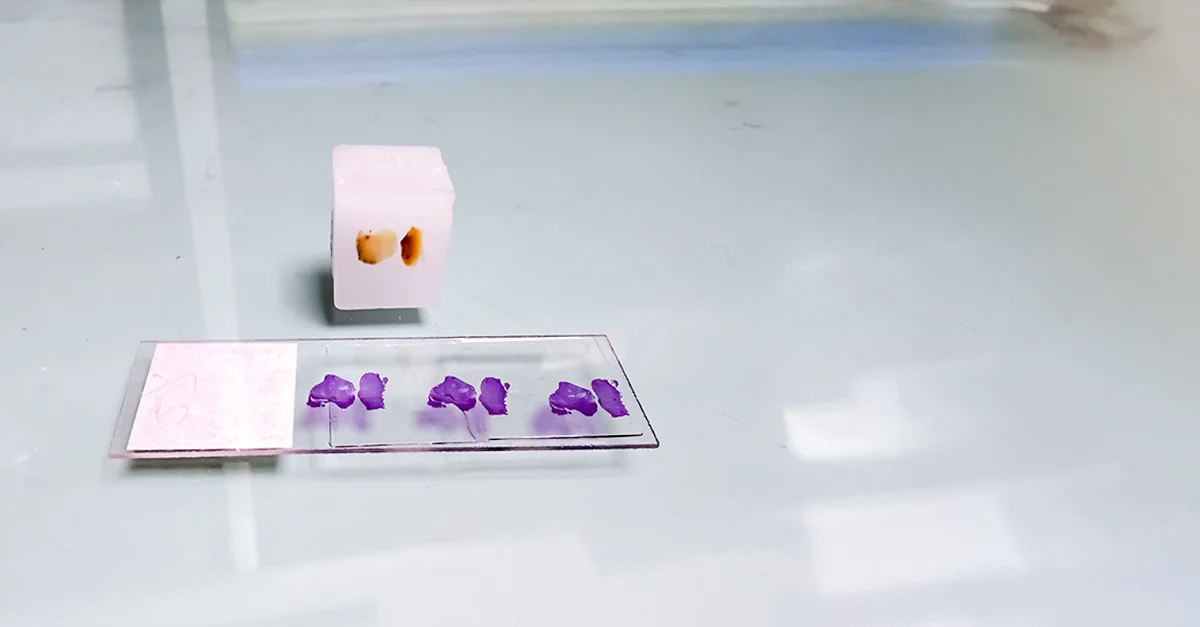
Mining formalin-fixed, paraffin-embedded (FFPE) samples represents one of, if not the, the biggest opportunity in translational biomedical research, because FFPE captures real-world disease biology at scale: fixation preserves tissue architecture and cellular detail, and embedding enables thin sectioning for staining and immunohistochemistry (IHC) (2,4). FFPE samples are also uniquely stable at room temperature for decades, if not longer (5,13,14), a feature that led FFPE to become and remains the default format of sample preservation in clinical pathology and biobanking (11,13,14). Resultingly, pathology and biobanking archives have grown to absolutely enormous scale – “billions” of FFPE specimens in published estimates (13,14) – sampled at literally every condition of health and disease at every stage (11); these samples are often paired with rich clinical annotation and long follow-up, and rare diseases and diverse treatment histories are well represented. To my knowledge, no other specimen types capture real-world human disease biology at a comparable scale, and no other sample is as physically robust. Yet the same stability and chemistry that makes FFPE so useful for histology creates challenges. Namely, wax and crosslinking are mutually incompatible with most analytical techniques, including proteomics. Hydrophobic paraffin contaminates, clogs column and results in unacceptable LC–MS performance (4,5,10). Formaldehyde fixation can cause chemical artifacts and literally turns samples into one giant molecule: proteins become locked into insoluble, inflexible networks that result in inefficient extraction, huge pellets and inhibited digestion. Unfortunately, “tissue in” often turns into “few peptides out” (3–6). Effective analysis to reveal the true underlying state of biology obligates that we rewind the very features that make tissue archival possible. However, FFPE proteomics does not have to be “second class.” With the right sample preparation including steps of extraction, homogenization and cleanup strategy, quantitative results from archival FFPE closely mirror that of paired flash-frozen tissues. Our HYPERsol workflow is a clear example: direct solubilization in 5% SDS, ultra- or megasonication (respectively at 500 kHz or 2 MHz) paired with S-Trap™ processing, yields depth and reproducibility on par with paired frozen tissue. Proteome quantifications also track with an average correlation of R = 0.94 and successful analysis of specimens stored for up to 17 years (5). Brief history of FFPE FFPE emerged when chemical sample fixation and paraffin infiltration embedding were efficiently combined into a single protocol (1,2,16,17). In the 1860s, paraffin infiltration embedding, in which water in tissue is removed by dehydration and clearing, then replaced with wax, was developed (16). Building on wax-based approaches described by Salomon Stricker (1834–1898) and paraffin experiments by Theodor Albrecht Edwin Klebs (1834–1913) that revealed challenges of infiltration, Wilhelm His Sr. (1831–1904) formalized a dehydration-clearing-paraffin infiltration (embedding) workflow that underlies modern practice (16). Fixing techniques were still evolving and in 1893, Ferdinand Blum (1865–1959), after noticing hardening of his fingertips during [ungloved] sample handling, showed that dilute formaldehyde “formalin” could preserve tissue with relatively little distortion while maintaining microscopic detail, making fixation reliable enough to withstand subsequent solvent and hot wax steps (2,17). This sequence defines the process we use today: [...]

By |2026-02-04T21:03:10+00:00Feb 04, 2026|Article|0 Comments
Go to Top